These Skin Bumps Are Not Pimples: Acne Mimicking Skin Conditions
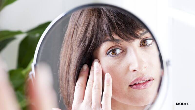
Due to the widespread occurrence of acne, many individuals often mistake any skin bump for a pimple or acne breakout. There are, however, numerous skin conditions that mimic acne but are distinctly different (and most importantly, they must be treated differently to effectively address or manage the condition).
Issues such as peri-orificial dermatitis, rosacea, and folliculitis are commonly mistaken for acne and as such they are not treated effectively. Accurate diagnosis at the onset of any skin change is critical. In this blog post, we will examine some common acne-like conditions and shed light on their causes, symptoms, similarities to acne, and appropriate treatments.
Folliculitis
Folliculitis is a common skin condition characterized by inflammation of hair follicles. When these follicles become irritated or infected, it leads to the development of red, swollen bumps or pustules. Folliculitis can occur anywhere on the body where hair grows, including the scalp, face, neck, chest, back, buttocks, and legs.
Symptoms of folliculitis may include:
- Red or inflamed skin around hair follicles
- Small red bumps or pustules
- Itching or burning sensation
- Tenderness or pain
- Formation of crusts or scabs (in more severe cases)
The treatment for folliculitis depends on the underlying cause and severity of the condition. Mild cases may resolve on their own or with self-care measures such as warm compresses and gentle cleansing. However, more severe or recurrent cases may require medical intervention, including antibiotics or topical creams.
In some cases, folliculitis may lead to complications such as furuncles (boils) or carbuncles (clusters of boils), which may require drainage or more aggressive treatment.
Keratosis Pilaris
Keratosis pilaris (KP) is a common, benign skin condition characterized by small, rough bumps on the skin’s surface. These bumps typically appear on areas of the body where hair follicles are most prominent, such as the upper arms, thighs, buttocks, and sometimes the face. Often referred to as “chicken skin,” keratosis pilaris is more noticeable when the skin is dry and tends to worsen during the winter months.
Symptoms of keratosis pilaris may include:
- Small, rough bumps on the skin, often described as resembling goosebumps or sandpaper
- Redness or inflammation around the bumps
- Itching or discomfort, especially in dry or cold weather
- Worsening of symptoms with friction or irritation, such as from clothing or shaving
Treatment options for keratosis pilaris may include:
- Moisturizers: Regular use of moisturizing creams or lotions can help soften the skin and reduce dryness, making the bumps less noticeable.
- Exfoliation: Gentle exfoliation with products containing alpha hydroxy acids (AHAs) or beta hydroxy acids (BHAs) can help remove dead skin cells and unclog the hair follicles.
- Topical retinoids: Prescription-strength retinoid creams or lotions can help promote cell turnover and prevent the formation of new keratin plugs.
- Laser therapy: Intense pulsed light (IPL) or fractional laser resurfacing may be used to improve the appearance of the skin and reduce redness.
Rosacea
Rosacea is a chronic inflammatory skin condition that primarily affects the face, causing redness, visible blood vessels, and acne-like bumps. It is a common condition that typically develops in adulthood, often between the ages of 30 and 50, though it can occur at any age. The cause of rosacea is believed to involve a combination of genetic, environmental, and vascular factors.
The symptoms of rosacea can vary widely from person to person but often include:
- Persistent facial redness: Rosacea commonly presents as redness or flushing on the central face, including the cheeks, nose, chin, and forehead. This redness may come and go or become more persistent over time.
- Visible blood vessels: Individuals with rosacea may develop visible blood vessels (telangiectasia) on the face, particularly around the nose and cheeks.
- Acne-like bumps: Some people with rosacea experience small, red bumps or pus-filled pimples resembling acne. These bumps may be accompanied by burning or stinging sensations.
- Thickened skin: In severe cases of rosacea, the skin on the nose may become thickened and enlarged, a condition known as rhinophyma.
- Eye symptoms: Ocular rosacea can cause irritation, redness, dryness, and sensitivity to light in the eyes. In some cases, untreated ocular rosacea can lead to more serious complications, such as corneal damage.
Treatment options for rosacea may include:
- Topical medications: Prescription creams or gels containing antibiotics, azelaic acid, or other anti-inflammatory agents may help reduce redness and inflammation.
- Oral antibiotics: Oral antibiotics may be prescribed to control inflammation and reduce acne-like bumps.
- Laser therapy: Laser or light-based treatments can help reduce redness, visible blood vessels, and thickened skin associated with rosacea.
- Avoidance of triggers: Identifying and avoiding triggers that exacerbate rosacea symptoms can help minimize flare-ups and improve the overall management of the condition.
- Skincare routine: Using mild cleansers and moisturizers, and avoiding harsh products and abrasive exfoliants, can help maintain the skin’s barrier function and reduce irritation.
- Eye care: Individuals with ocular rosacea may benefit from lubricating eye drops, warm compresses, and prescription medications to manage eye symptoms.
Syringoma
Syringoma is a benign skin condition characterized by small, firm bumps that typically appear on the face, particularly around the eyes, cheeks, and forehead. These bumps are usually skin-colored or yellowish and develop from the eccrine glands (sweat glands).
Syringomas are typically diagnosed based on their appearance, though a biopsy may be performed to confirm the diagnosis. These bumps are often mistaken for acne or milia, but unlike acne, they are not associated with inflammation or pus.
If treatment is needed, several options may be considered:
- Topical treatments: Certain topical medications, such as retinoids or corticosteroids, may help reduce the size or appearance of syringomas over time.
- Electrocautery: Also known as electrosurgery, this treatment involves using a small electrical probe to heat and destroy the syringoma tissue. This procedure is typically performed by a dermatologist and may require multiple sessions for optimal results.
- Laser therapy: Laser treatments can be effective in reducing the size and appearance of syringomas.
- Cryotherapy: Cryotherapy involves freezing the syringomas with liquid nitrogen to destroy the abnormal tissue. This procedure may cause temporary redness, swelling, and blistering, but the skin typically heals within a few weeks.
- Surgical excision: In some cases, particularly for larger or more bothersome syringomas, surgical excision may be recommended. This involves cutting out the syringoma tissue under local anesthesia and suturing the skin closed.
Peri-Orificial Dermatitis
Peri-orificial dermatitis is a common inflammatory skin condition that typically affects the area around the mouth, nose, and eyes. It is characterized by small, red bumps or pustules that may be accompanied by mild itching or burning sensations. Perioral dermatitis primarily affects women between the ages of 20 and 45, although it can occur in men and children as well.
Peri-orificial dermatitis can be a result of a combination of factors, including:
- Topical steroids: Long-term use of topical steroids, such as corticosteroid creams or ointments, on the face can trigger or exacerbate perioral dermatitis. This is known as steroid-induced perioral dermatitis.
- Fluoride toothpaste: Some studies suggest that fluoride toothpaste may contribute to the development of perioral dermatitis, particularly in individuals who are sensitive to fluoride.
- Skin irritants: Harsh skincare products, cosmetics, or facial cleansers containing fragrances, dyes, or other irritants may irritate the skin and contribute to the development of perioral dermatitis.
- Hormonal factors: Fluctuations in hormone levels, such as those occurring during pregnancy, menstruation, or menopause, may play a role in the development of perioral dermatitis.
Symptoms of peri-orificial dermatitis may include:
- Small, red bumps or pustules around the mouth, nose, or eyes
- Mild itching or burning sensations
- Dry or flaky skin
- Redness or inflammation
- Occasionally, the bumps may ooze or crust over
Depending on the severity of the condition, treatment options may include:
- Topical antibiotics: Prescription-strength antibiotics may be applied topically to reduce inflammation and kill bacteria on the skin.
- Oral antibiotics: In more severe cases, oral antibiotics may be prescribed to help control inflammation and prevent recurrence.
- Topical immunosuppressants: In cases where other treatments have been ineffective, topical calcineurin inhibitors may be used to reduce inflammation and suppress the immune response.
- Gentle skincare: Using mild, fragrance-free cleansers and moisturizers can help soothe the skin and prevent further irritation. Avoiding harsh skincare products or cosmetics that may exacerbate symptoms is also important.
- Lifestyle modifications: Making lifestyle changes, such as avoiding spicy foods, alcohol, and excessive sun exposure, may help reduce flare-ups of perioral dermatitis.
Staph Bacteria Infection
Commonly referred to as a staph infection, staphylococcus bacteria are ubiquitous and can be found on the skin and mucous membranes of healthy individuals. While they typically do not cause harm under normal circumstances, they can cause infections when they enter the body through cuts, wounds, or other breaks in the skin.
There are several types of staph infections, ranging from minor skin infections to more severe, life-threatening conditions. Staph bacteria can cause various skin infections, such as boils, impetigo (a highly contagious skin infection), cellulitis (a bacterial infection of the deeper layers of the skin), and folliculitis (inflammation of hair follicles, mentioned above). These infections typically present as red, swollen, painful areas on the skin and may be accompanied by pus or drainage.
Treatment for staph infections may involve:
- Antibiotics: Depending on the severity and type of infection, antibiotics may be prescribed to kill the bacteria and prevent further spread of the infection. In severe cases, intravenous antibiotics may be necessary.
- Incision and drainage: A healthcare provider may need to drain the infected fluid to promote healing and relieve symptoms.
- Wound care: Proper wound care can help prevent staph infections and promote healing.
- Hospital care: In cases of severe infections, care in a hospital setting may be necessary to monitor vital signs, administer fluids and medications, and provide other supportive measures.
Sebaceous Hyperplasia
Sebaceous hyperplasia is a common benign skin condition characterized by the enlargement of sebaceous glands. Sebaceous hyperplasia typically presents as small, flesh-colored or yellowish bumps on the skin, particularly on the face, especially on the forehead, cheeks, and nose.
The exact cause of sebaceous hyperplasia is believed to result from a combination of genetic factors, hormonal influences, and aging. Sebaceous glands may become enlarged or overactive due to hormonal changes, such as those occurring during puberty, pregnancy, or menopause. Additionally, sun exposure and damage to the skin may contribute to the development of sebaceous hyperplasia.
Treatment for sebaceous hyperplasia may include:
- Topical treatments: Certain topical medications, such as retinoids or salicylic acid, may help reduce the size and appearance of sebaceous hyperplasia bumps over time by promoting cell turnover and exfoliation of the skin.
- Electrocautery: Electrocautery involves using a small electrical probe to heat and destroy the enlarged sebaceous glands.
- Cryotherapy: Cryotherapy is performed to freeze the sebaceous hyperplasia bumps with liquid nitrogen to destroy the overactive sebaceous glands.
- Laser therapy: Laser treatments can be effective in reducing the size and appearance of sebaceous hyperplasia bumps.
- Surgical excision: In some cases, particularly for larger or more bothersome bumps, surgical excision may be recommended.
Steatocystoma Multiplex
Steatocystoma multiplex is a rare genetic skin disorder characterized by the development of multiple cysts filled with oily or cheesy material beneath the skin. These cysts typically appear on the upper body, particularly the chest, neck, upper arms, and back, but they can also occur on the face and scalp.
The cause of steatocystoma multiplex is believed to be a result of abnormalities in the development of sebaceous glands. In individuals with steatocystoma multiplex, the ducts of the sebaceous glands become blocked, leading to the accumulation of sebum and the formation of cysts beneath the skin’s surface. In many cases, treatment is not necessary unless the cysts are causing symptoms or cosmetic concerns.
However, if treatment is desired, options may include:
- Incision and drainage: A healthcare provider may perform a minor surgical procedure to drain the cyst and relieve symptoms.
- Cyst excision: In some cases, particularly for larger or more bothersome cysts, surgical excision may be recommended.
- Laser therapy: Laser treatments can be effective in reducing the size and appearance of cysts.
- Intralesional injections: Intralesional injections of corticosteroids may be used to reduce inflammation and shrink cysts in some cases.
Basal Cell Carcinoma
Basal cell carcinoma (BCC) is the most common type of skin cancer, accounting for about 80% of all cases of skin cancer. It typically develops in areas of the skin that are frequently exposed to the sun, such as the face, neck, scalp, and arms, although it can occur in other areas as well. Basal cell carcinoma tends to grow slowly and is usually not life-threatening, but it can cause significant damage if left untreated.
The cause of basal cell carcinoma is believed to be primarily caused by long-term exposure to ultraviolet (UV) radiation from the sun or tanning beds.
Other risk factors for basal cell carcinoma include:
- Fair skin: People with fair skin, light hair, and blue or green eyes are at higher risk of developing basal cell carcinoma because they have less natural protection against UV radiation.
- Chronic sun exposure: Spending a lot of time outdoors without sunscreen or protective clothing increases the risk of developing basal cell carcinoma.
- History of sunburns: Having a history of sunburns, particularly during childhood or adolescence, increases the risk of developing basal cell carcinoma later in life.
- Personal or family history: People who have previously had basal cell carcinoma or other types of skin cancer, as well as those with a family history of skin cancer, are at increased risk.
Basal cell carcinoma usually appears as a pearly or waxy bump on the skin, often with visible blood vessels. It may also present as a flat, flesh-colored or brownish patch that gradually enlarges over time.
Other possible signs of basal cell carcinoma include:
- A sore that does not heal
- A shiny, translucent bump
- A red, scaly patch
- Pink growths with raised borders
- Open sores that may ooze or crust over
Once diagnosed, treatment options for basal cell carcinoma may include:
- Surgical excision: Surgical removal of the tumor is often the preferred treatment for basal cell carcinoma. The surgeon removes the tumor along with a margin of healthy skin to ensure complete removal of cancerous cells.
- Mohs surgery: Mohs micrographic surgery is a specialized technique that involves removing thin layers of skin one at a time and examining them under a microscope until no cancer cells are detected. Mohs surgery is often used for tumors in cosmetically sensitive areas or those with unclear margins.
- Electrodessication and curettage: This procedure involves scraping away the cancerous tissue and then cauterizing the area with an electric current to destroy any remaining cancer cells.
- Cryotherapy: Cryotherapy involves freezing the cancerous tissue with liquid nitrogen, causing it to blister and eventually slough off.
- Topical medications: Topical medications may be prescribed to treat superficial basal cell carcinomas or lesions in difficult-to-treat areas.
Molluscum Contagiosum
Molluscum contagiosum is a common viral skin infection that causes small, raised bumps or lesions on the skin. It is caused by the molluscum contagiosum virus (MCV), a member of the poxvirus family. Molluscum contagiosum is highly contagious and is spread through direct skin-to-skin contact with an infected person or by sharing items such as towels or clothing.
The characteristic symptom of molluscum contagiosum is the development of small, flesh-colored, or pearly-white bumps on the skin. These bumps are often dome-shaped and may have a small indentation or dimple in the center.
Molluscum contagiosum lesions can appear anywhere on the body but are most found on the face, neck, arms, hands, and genital area. Treatment may be recommended for individuals with persistent or bothersome symptoms, especially if the lesions are in sensitive areas or if there is a risk of spreading the infection to others.
Treatment options for molluscum contagiosum may include:
- Topical treatments: Certain topical medications, such as imiquimod cream or tretinoin cream, may be prescribed to help stimulate the immune system and promote the clearance of the virus.
- Cryotherapy: Cryotherapy is performed to freeze the lesions with liquid nitrogen to destroy the infected tissue.
- Curettage: Curettage involves scraping the lesions off the skin. This procedure is often performed in conjunction with cryotherapy to remove the remaining tissue.
- Laser therapy: Laser treatments can be effective in removing molluscum lesions while minimizing scarring and pigmentation changes.
- Home care: Over-the-counter remedies may help reduce the size and appearance of molluscum lesions.
Milia
Milia are small, benign cysts that commonly appear on the skin as tiny white or yellowish bumps. These cysts form when keratin, a protein found in the skin, becomes trapped beneath the surface, leading to the formation of small, raised bumps. Milia is commonly found on the face, particularly around the eyes, cheeks, nose, and forehead. However, they can also occur on other parts of the body.
While milia are harmless and typically resolve on their own over time, some people may choose to seek treatment to improve their appearance or prevent recurrence.
Treatment options for milia may include:
- Exfoliation: Gentle exfoliation with a mild facial scrub or chemical exfoliant containing ingredients such as AHAs or BHAs can help remove dead skin cells and promote the shedding of milia.
- Topical retinoids: Prescription-strength retinoid creams or gels containing ingredients such as tretinoin or adapalene may help promote cell turnover and prevent the formation of new milia.
- Extraction: A dermatologist or skincare professional may use a sterile needle or lancet to carefully extract the contents of milia. This procedure should only be performed by a trained professional to minimize the risk of scarring or infection.
- Laser therapy: Laser treatments can be effective in removing milia by targeting and destroying the cysts.
While acne is a prevalent skin concern, it is essential to recognize that not all skin bumps are pimples. Various conditions mimic acne but require distinct approaches to management. Accurate diagnosis by a dermatologist ensures appropriate treatment, alleviating symptoms and preventing potential complications. By understanding the differences between acne and its mimics, patients can make informed decisions about their skincare and seek medical attention when needed.
Dr. Neil Farnsworth is a board-certified dermatologist with longstanding ties to the Houston area. Dr. Farnsworth believes in a comprehensive approach to dermatology, with a special emphasis on regular maintenance of the skin’s health through sun-protection, use of the proper personal care products, and vigilance against potential assaults such as infections, skin cancers, and autoimmune disease. Dr. Farnsworth sees patients at our River Oaks location.
Disclaimer: The contents of the Westlake Dermatology website, including text, graphics, and images, are for informational purposes only and are not intended to substitute for direct medical advice from your physician or other qualified professional.
I have tiny perfectly shaped hard bumps on my legs . They are solid white and hard